Artificial Intelligence in Radiology in Partnership with Gleamer
Visida Solutions is the official partner of Gleamer in Latvia, providing advanced artificial intelligence solutions for radiology to healthcare institutions and diagnostic centers.
Gleamer is a leading French medical AI company developing CE-marked and clinically validated AI solutions for medical imaging. Its technologies support radiologists in increasing diagnostic accuracy, improving workflow efficiency, and enhancing clinical decision-making.
X-Ray Solutions
BoneView
AI-Powered Trauma X-ray Interpretation
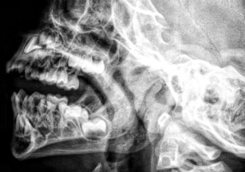
BoneView is an artificial intelligence solution designed to support radiologists and emergency physicians in the detection of bone fractures and skeletal injuries on standard X-ray images.
Real time radiograph analysis
The system analyzes radiographs in real time, highlighting suspicious areas and potential fractures to assist clinicians in making faster and more confident diagnostic decisions, especially in time-critical trauma and emergency settings.
Enhanced diagnostic accuracy
BoneView improves diagnostic efficiency by reducing the risk of missed findings and helping radiologists prioritize complex cases. It supports consistent interpretation across different experience levels and helps decrease reporting time without changing the existing clinical workflow.
Thorough validation & training
The solution has been clinically validated and trained on large and diverse datasets covering a wide range of skeletal anatomies and fracture types. It integrates seamlessly with PACS and radiology systems, providing AI results directly in the radiologist’s reading environment.
Valuable in rapid environments
BoneView is particularly valuable in high-volume trauma centers and emergency departments, where rapid, accurate triage and interpretation of X-ray studies are essential for patient care.
BoneMetrics
AI-Powered Bone Age Assessment
BoneMetrics is an advanced artificial intelligence solution for automated pediatric bone age assessment based on hand and wrist X-ray imaging.
Fast and reliable estimations
The system analyzes radiographic images in seconds, providing fast, consistent, and clinically reliable bone age estimations. By standardizing the assessment process, BoneMetrics significantly reduces inter-observer variability and minimizes manual effort for radiologists and pediatric specialists.
Thorough validation & training
BoneMetrics is trained on large, diverse pediatric datasets and clinically validated across multiple populations and imaging conditions. It supports both routine clinical practice and specialized pediatric endocrinology workflows, helping clinicians monitor growth disorders, assess developmental delays, and support treatment planning.
Increased workflow efficiency
By reducing reporting time and improving reproducibility, BoneMetrics increases workflow efficiency while maintaining a high level of diagnostic quality. The solution integrates seamlessly into existing PACS and radiology information systems without disrupting daily clinical routines.
BoneAge
Automated Bone Age Assessment in Seconds
BoneAge is an AI-powered solution for fast and accurate pediatric bone age assessment. It automatically analyzes hand and wrist X-ray images and delivers results within seconds, significantly reducing manual workload for radiologists and pediatric specialists.
Thorough validation & training
BoneMetrics is trained on large, diverse pediatric datasets and clinically validated across multiple populations and imaging conditions. It supports both routine clinical practice and specialized pediatric endocrinology workflows, helping clinicians monitor growth disorders, assess developmental delays, and support treatment planning.
Seamless integration
It integrates seamlessly into existing radiology workflows without changing routine clinical practice.
ChestView
AI-Powered Chest X-ray Analysis
ChestView is an artificial intelligence solution designed to support radiologists in the detection of thoracic and pulmonary abnormalities on chest X-ray images.
Real time radiograph analysis
The system analyzes chest radiographs in real time and highlights potential pathological findings, such as lung opacities and other clinically relevant changes. This helps radiologists improve interpretation speed and maintain a high level of diagnostic consistency, particularly in high-volume environments such as emergency departments and outpatient radiology centers.
Thorough validation & training
ChestView is trained on large, diverse datasets and clinically validated to perform across different patient populations, imaging conditions, and equipment vendors. It supports early identification of abnormalities, helping clinicians prioritize urgent cases and reduce the risk of overlooked findings.
Seamless integration without disruption
By integrating directly into the existing PACS and radiology workflow, ChestView provides AI assistance without disrupting routine clinical processes. It enables radiologists to work more efficiently while maintaining full control over the final diagnosis and clinical decision-making.
Valuable in high-volume environments
ChestView is especially valuable for institutions handling large volumes of chest X-ray examinations, supporting faster triage, improved workflow efficiency, and more standardized reporting.
BreastView
AI-Powered Chest X-ray Analysis
BreastView is an advanced artificial intelligence solution for mammography and digital breast tomosynthesis, designed to support radiologists in breast cancer screening and diagnostic workflows.
Automatic analysis of breast imaging
The system automatically analyzes breast imaging studies and assists in identifying suspicious lesions by highlighting areas of interest directly on the images. It also provides breast density classification according to the BI-RADS A–D standard, helping support risk stratification and clinical reporting.
Thorough validation & training
BreastView delivers malignancy risk assessment using standardized AI metrics, supporting radiologists in prioritizing cases and improving diagnostic confidence. The solution is clinically validated on large, diverse datasets and developed to perform reliably across different patient populations and imaging equipment.
Increased efficiency & consistency
By reducing visual fatigue and helping detect subtle findings, BreastView enhances both efficiency and consistency in breast imaging interpretation. It integrates seamlessly into existing PACS and radiology workflows, allowing radiologists to benefit from AI support without changing their daily practice or reporting process.
Valuable in high-volume environments
BreastView is particularly valuable for breast cancer screening programs and high-volume diagnostic centers, where accuracy, standardization, and efficiency are critical for improving patient outcomes.
Computed tomography
LungCT
AI-Powered Lung Nodule Detection and Analysis
LungCT is an advanced artificial intelligence solution for automated lung nodule detection and analysis on chest CT examinations, designed to support radiologists in lung cancer screening and longitudinal patient monitoring.
Automated lung nodule detection
The system automatically identifies lung nodules, provides precise measurements, and tracks changes in size and volume over time. It also offers an AI-based malignancy risk assessment to help radiologists prioritize findings and standardize clinical evaluation across different users and institutions.
Thorough validation & training
LungCT has been trained on large, multi-institutional datasets and clinically validated to ensure robust performance across diverse patient populations, CT scanners, and imaging protocols. It supports consistent nodule characterization and follow-up, improving both diagnostic accuracy and workflow efficiency.
Reduced manual workload
By reducing manual measurement time and inter-reader variability, LungCT helps radiologists focus on clinical interpretation while ensuring structured and reproducible assessment of pulmonary nodules. Its seamless integration with existing PACS and radiology systems allows AI insights to be available directly within routine reporting workflows.
Valuable in cancer screening processes
LungCT is particularly valuable in lung cancer screening programs and high-throughput imaging centers, where early detection and standardized nodule management are critical to improving patient outcomes.
BoneCT
AI-Powered Bone Lesion Detection on CT
BoneCT is the world’s first fully developed artificial intelligence solution dedicated to the detection and characterization of bone lesions on CT imaging.
Automated bone lesion detection
The system automatically identifies both sclerotic and osteolytic bone lesions, supporting radiologists in the assessment of primary bone tumors, metastatic disease, and other pathological bone changes. By analyzing complex bone structures across CT datasets, BoneCT assists in early lesion detection, classification, and prioritization of clinically relevant findings.
Thorough validation & training
BoneCT is trained on large, diverse CT datasets and clinically validated to perform across different scanners, patient populations, and anatomical regions. It supports radiologists in identifying subtle or easily overlooked bone changes, especially in high-volume oncology and trauma imaging workflows.
Enhanced diagnostic accuracy
By reducing manual review time and improving detection consistency, BoneCT helps enhance diagnostic accuracy while maintaining compatibility with existing clinical workflows. The solution integrates seamlessly with PACS and radiology systems, allowing AI outputs to be accessed directly in the radiologist’s reading environment.
Valuable in precision heavy environments
BoneCT is particularly valuable in oncological imaging, musculoskeletal radiology, and trauma diagnostics, where precise evaluation of bone lesions is critical for patient management and treatment planning.
Magnetic Resonance
NeuroMR
AI-Powered Brain MRI Analysis
NeuroMR is an advanced artificial intelligence solution for automated analysis of brain MRI studies, designed to support radiologists and neurologists in the assessment and monitoring of neurological diseases.
Automated brain MRI analysis
The system provides accurate detection and quantification of multiple sclerosis (MS) lesions, enabling objective evaluation of lesion burden and disease progression over time. In addition, NeuroMR delivers detailed brain volumetric analysis, supporting the assessment of brain atrophy and structural changes associated with various neurological conditions.
Thorough validation & training
NeuroMR is trained on large, multi-center MRI datasets and clinically validated to ensure reliable performance across different MRI scanners, protocols, and patient populations. It supports longitudinal analysis by enabling consistent follow-up comparisons between current and prior examinations, which is essential for treatment monitoring and therapy evaluation.
Complex process automation
By automating complex and time-consuming measurements, NeuroMR reduces manual workload and inter-observer variability, while improving reproducibility and standardization in neuroradiological reporting. The solution integrates seamlessly into existing PACS and radiology workflows, allowing radiologists to access AI-generated insights directly within their normal reporting environment.
Valuable insights and precision
NeuroMR is particularly valuable in multiple sclerosis management, neurodegenerative disease assessment, and research environments requiring precise, standardized brain MRI analysis.
LumbarMR
AI-Powered Lumbar Spine MRI Analysis
To be launched soon
LumbarMR is an artificial intelligence solution currently under development for automated analysis of lumbar spine MRI studies, designed to support the detection and assessment of common spinal pathologies.
Improved diagnostic efficiency
The solution focuses on identifying and characterizing degenerative changes, intervertebral disc pathology, spinal canal stenosis, and other clinically relevant lumbar spine abnormalities. By assisting radiologists in evaluating complex spinal structures, LumbarMR aims to improve diagnostic efficiency and reduce manual interpretation time.
Thorough development & testing
LumbarMR is being developed using diverse, high-quality MRI datasets and leading AI methodologies to ensure robust performance across different scanners, imaging protocols, and patient populations. The solution is intended to support standardized reporting and longitudinal monitoring of spinal conditions over time.
Seamless integration
Once released, LumbarMR will integrate seamlessly into existing PACS and radiology workflows, providing AI-generated insights directly within the radiologist’s reading environment without disrupting routine clinical practice.
Valuable in high volume environments
LumbarMR is expected to be particularly valuable for radiology departments managing high volumes of spine MRI examinations, supporting both routine diagnostics and specialized musculoskeletal imaging workflows.
Get in touch at
info@visidasolutions.com